FIGURE 22.
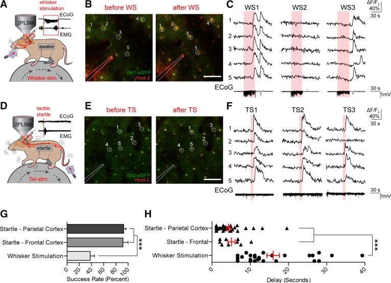
Startle stimulation triggers widespread astroglial Ca2+ signaling. A: astrocytic Ca2+ transients measured by rhod 2-AM were detected in response to 30 s of 3-Hz whisker stimulation. Representative local field potential LFP and electromyogram (EMG) recordings are shown. B: representative images of rhod 2-AM fluorescence increases during whisker stimulation in a Glt-1-EGFP animal. Scale bar = 100 μM. C: selected cells from B. Rhod 2 ΔF/F0 was normalized to Glt-1-EGFP fluorescence. Electrocorticogram (ECoG) traces corresponding to rhod 2 ΔF/F0 are shown below. D: air pulses were directed at the face or tail of the animal to elicit a startle response. Representative ECoG and EMG traces are shown with no apparent evoked ECoG response and strong EMG activity, indicative of a startle response. E and F: representative images and corresponding rhod 2 ΔF/F0 traces show stable and repeatable astrocytic [Ca2+]i transients after startle stimulations. Bottom: representative ECoG traces from startle stimulation are shown. G: bar graph showing the response rate of cortical astrocytes to whisker and startle stimulation, averaged across animals. *** P < 0.001, one-way ANOVA, Bonferroni correction [n = 11 animals (parietal cortex startle and whisker stimulation), 4 animals (frontal cortex startle)]. H: scatter diagram with superimposed mean and SE for the delay from the onset of whisker/startle stimulation to the beginning of astrocytic rhod 2 ΔF/F0 responses. Average delay from each successful trial is shown. *** P < 0.001, one-way ANOVA, Bonferroni correction [n = 28 trials in 14 animals (whisker stimulation), 38 trials in 13 animals (parietal cortex startle), and 9 trials in 4 animals (frontal cortex startle)]. Data are means ± SE. [From Ding et al. (431).]

