FIGURE 30.
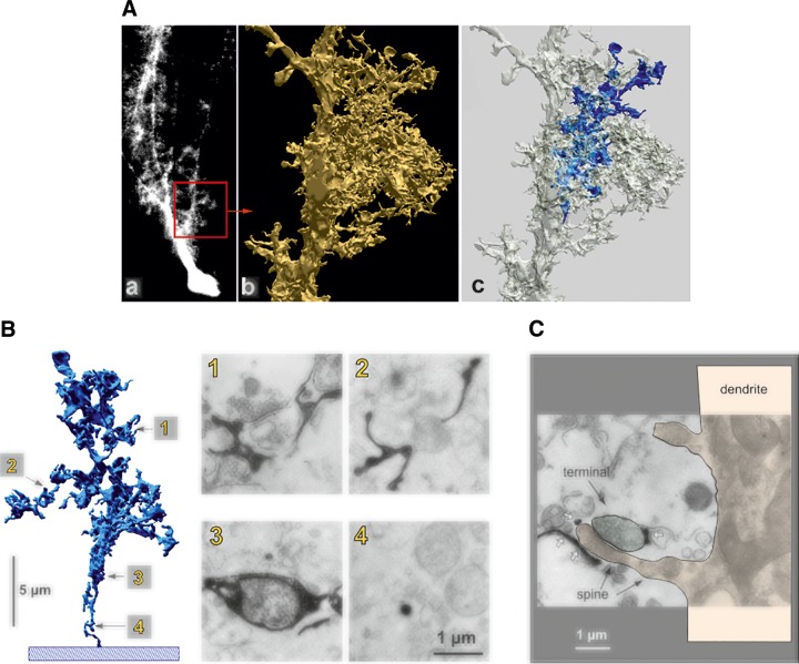
Microdomain organization of Bergmann glia and coverage of parallel fibers to Purkinje neuron synapses. A: reconstruction of an appendage based on electron microscopic data. a: Fluorescence light micrograph of a dye-injected Bergmann glial cell is shown; the red square (20 × 20 mm) corresponds to the portion that was reconstructed from consecutive ultrathin sections. b: One of the lateral appendages, arising directly from fiber with all the other side branches omitted for clarity. c: The same appendage as shown in b, but with one of the appendages marked by blue. This labeled structure is shown in isolation and at higher magnification in B. B: fine structure of appendages and relation to synapses. a: A small lateral appendage, arising from the reconstructed part of the glial fiber (blue in Ac), is shown as a slightly turned, isolated three-dimensional reconstruction (left). Electron micrographs of four sections contributing to the reconstruction (designated 1–4) are shown on the right; glial compartments appear black after conversion of the injected dye. The location of these sections in the reconstruction is indicated by the labeled arrows: 1, region directly contacting synapses; 2, glial compartments without direct synaptic contacts; 3, bulging glial structure containing a mitochondrion; 4, the stalk of the appendage. C: another example of a synapse contacted by glial compartments, appearing as black structures (white arrows); in this case, the postsynaptic element can be traced back along the spine (black arrows) to the Purkinje cell dendrite (reddish overlay; the presynaptic terminal is labeled by a green overlay). [From Grosche et al. (609).]

