
FIGURE 2.
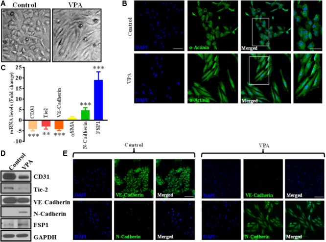
Valproic acid causes marked morphological and ultrastructural changes, and promotes EndMT-like phenotypic switching in HUVECs. (A) Diluent-treated control HUVECs, cultured on a two-dimensional plate, formed a confluent monolayer with the typical EC ‘cobblestone’ morphology (left panel). VPA treatment resulted in marked morphological changes whereby HUVECs took on an enlarged spindle-shaped appearance with smooth surfaces (right panel). Both micrographs were taken at the same magnification (10X). (B) Immunofluorescent micrographs demonstrating cytoskeletal protein re-organization in HUVECs following VPA treatment. α-Actinin positivity is indicated in green and nuclei were stained with DAPI (blue); scale bar = 10 μm. (C) HUVECs were treated with diluent control or VPA. Total RNA and protein were extracted at 24 and 48 h, respectively. Differential (C) transcript (qPCR) ∗∗p < 0.01, ∗∗∗p < 0.001 and (D) protein (immunoblotting) levels of key endothelial and mesenchymal markers as well as (E) VE-Cadherin and N-Cadherin immunofluorescent staining in control- and VPA-treated HUVECs indicate EndMT with VPA treatment. Nuclei were stained with DAPI (blue). Micrographs are representative images of HUVECs taken 48 h post-treatment; scale bar = 20 μm.