FIGURE 4.

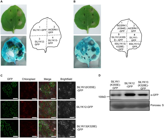
Overexpression of SlLYK1 and SlLYK13 induces cell death in Nicotiana benthamiana leaves. (A) Detection of cell death in N. benthamiana leaves. C terminus of AtCERK1, SlLYK1, SlLYK12, and SlLYK13 proteins were fused to green fluorescence protein (GFP) and then transiently expressed in N. benthamiana. Images were taken before (above) and after trypan blue staining (below) 3 days after infiltration. (B) Kinase inactivation of SlLYK1(K355E), SlLYK13(K328E), and AtCERK1(K394E) abolished the symptoms of cell death. (C) GFP observation. Images were taken using confocal microscopy 3 days after infiltration. Bars, 50 μM. (D) Size and abundance of the SlLYK1(K355E)-GFP, SlLYK12-GFP, and SlLYK13(K328E)-GFP proteins. Total proteins were extracted from leaves expressing the SlLYK1(K355E)-GFP, SlLYK12-GFP, and SlLYK13(K328E)-GFP constructs. Immunoblot analysis was performed using anti-GFP antibody. Ponceau S staining was used as a protein loading control. The experiment was repeated twice with similar results.
