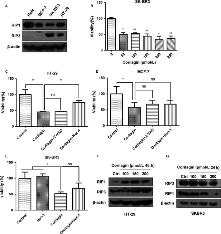
Figure 5.

Corilagin cannot induce necroptosis in SK‐BR3 cells. (A) The protein levels of RIP1 and RIP3 were detected by Western blot analysis in different cell lines. (B) SKBR3 cells were treated with 0, 50, 100, 150, 200, 250 μmol/L corilagin for 48 h. MTT assay was performed to assess the growth inhibiting effects. (C) HT‐29 cells and (D) MCF‐7 cells were pretreated with z‐VAD‐fmk and Nec‐1 for 2 h and then co‐treatment with corilagin for 24 h. Cell viability was detected by MTT assay. (E) SK‐BR3 cells pretreated with Nec‐1 for 2 h and then co‐treatment with 100 μmol/L corilagin for 48 h. Cell viability was detected by MTT assay. (F) HT‐29 cells and (G) SK‐BR3 cells were treated by corilagin. The expression of RIP3 and RIP1 was detected by Western blot. Data are expressed as means (n ≥ 3) ± SD over controls, *P < .05, **P < .01, ns: no significant. Abbreviations: Nec‐1, necrostatin‐1
