Figure 4.

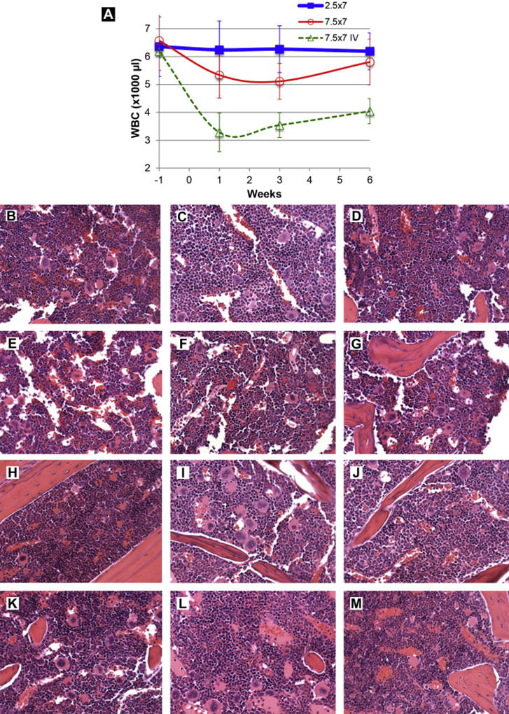
a. White blood cell count. Three groups of mice were treated once daily for 7 days by either aerosolized Aza at 2.5 mg/m2 (0.83 mg/kg; square) or 7.5 mg/m2 (2.5 mg/kg; round), or tail vein injection of Aza at 7.5 mg/m2 (2.5 mg/kg; triangle, dash line). Fifty microliter blood samples from each mouse were obtained under isoflurane anesthesia from the facial vein18 at 1 week prior and 1, 3, and 6 weeks after the treatment. White blood cells (WBCs) were collected and counted with a hemocytometer under a microscope after removing red blood cells. The data was mean and standard deviation from 6 to 12 mice per time point.
b~m. Pathological evaluation of the myelosuppression. Representative images of bone marrow (from sternum) 3 weeks (b~g) or 6 weeks (h~m) after the final administration of aerosolized Aza at 2.5 mg/m2 (0.83 mg/kg) daily × 7 days. In all cases, the bone marrow was histologically normal. The pictures were taken from hematoxylin and eosin stained slides and the objective magnification was 40×.
