Figure 5.

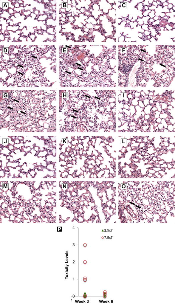
Pathological evaluation of the lungs from mice treated with aerosolized vehicle or Vidaza at 3 and 6 weeks after final treatment. a~c: Lungs from mice treated with aerosolized vehicle (10 mg/ml mannitol in sterile water). The lungs are normal. d~i: Lungs from mice treated with 7.5 mg/m2 (2.5 mg/kg) of aerosolized Vidazla three weeks after the final dose. There are low numbers of scattered foamy macrophages (arrows) within the alveoli in 5 of 6 mice. j~o: Lungs from mice treated with 7.5 mg/m2 (2.5 mg/kg) of aerosolized Vidazla six weeks after the final dose. One of the 6 mice had small alveolar macrophages (arrows), which was incidental and not a toxic lesion (i.e. occasionally present in vehicle-treated animals). The pictures were taken from hematoxylin and eosin stained slides and the objective magnification was 40×. p: The lung toxicity score: The toxicity of each lung were evaluated pathologically and characterized as 5 different grades: 0 = no toxicity/normal, 1 = minimal, 2 = moderate, 3 = median, 4 = severe. The circles represent high dose (7.5 mg/m2 or 2.5 mg/kg daily ×7, the H & E pictures presented above d~o), the solid triangles represent low dose (2.5 mg/m2 or 0.83 mg/kg daily ×7).
