Fig. 1.
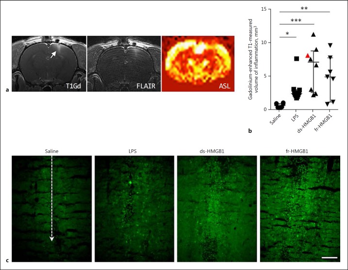
Intracerebral injection of HMGB1 induces blood-brain barrier leakage and neuroinflammation. HMGB1, high-mobility group box 1; LPS, lipopolysaccharide; ds, disulfide; fr, fully reduced. a Representative T1-weighted, FLAIR, and ASL images (non-tagged-tagged/non-tagged) collected after ds-HMGB1 injection (animal identified by a red triangle in b). T1-weighted and FLAIR images were collected after gadolinium. b Quantification of the volume of inflammation in gadolinium-enhanced T1-weighted images. LPS, ds-HMGB1, and fr-HMGB1 significantly increased gadolinium uptake compared to the saline controls (saline vs. LPS, p = 0.0413; saline vs. ds-HMGB1, p = 0.0002; saline vs. fr-HMGB1, p = 0.0076). Data are expressed as median ± IQR. p values were calculated using a Kruskal-Wallis test corrected for multiple comparisons using the Dunn test (n = 7–10 rats/group). * p ≤ 0.05, ** p ≤ 0.01, *** p ≤ 0.001. c Representative images showing extravasation of 70 kDa FITC-conjugated dextran. Dashed line indicates the location of the injection site (n = 3–4 rats/group). Scale bar, 100 µm.

