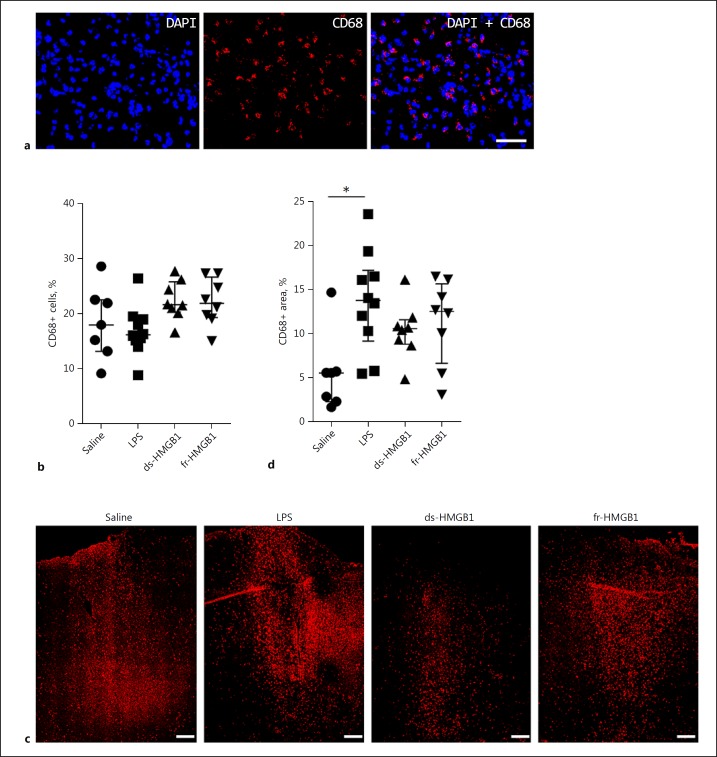
Fig. 2.
CD68 protein levels are increased in response to cerebral LPS but not HMGB1. Representative sections showing the level of CD68 protein present at the injection site and in the ipsilateral hemisphere. See legend to Fig. 1 for abbreviations. a High-magnification images showing CD68+ staining at the injection site. Scale bar, 25 µm. b Quantification of CD68+ cells at the injection site. No significant differences were detected between the groups. c Low magnification image illustrating the distribution of CD68+ cells in the cerebral cortex of the ipsilateral hemisphere. Scale bar, 200 µm. d Total area positive for CD68 cells in the ipsilateral hemisphere. Rats injected with LPS had increased CD68 protein levels compared to the saline controls (p = 0.0116). The difference in the HMGB1-treated animals was not significant. Data are expressed as median ± IQR. p values were calculated using a Kruskal-Wallis test corrected for multiple comparisons using the Dunn test (n = 8–10 rats/group). * p ≤ 0.05, ** p ≤ 0.01, *** p ≤ 0.001.

