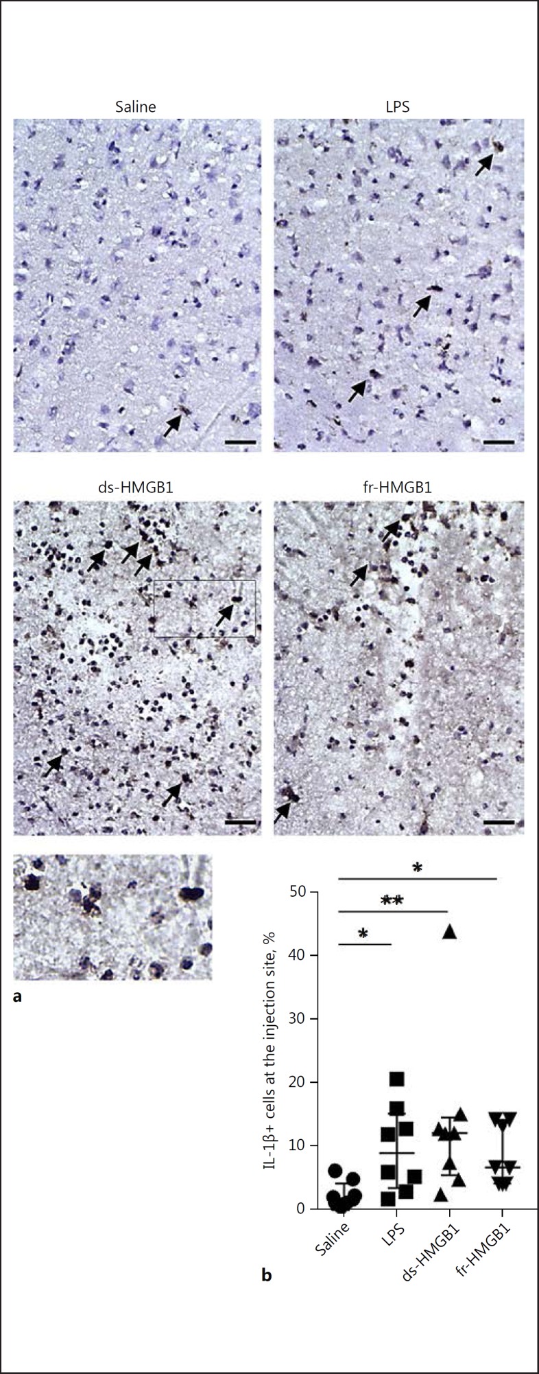
Fig. 4.
Intracerebral extracellular HMGB1 and LPS induce proinflammatory cytokine production. a Representative sections stained using an anti-IL-1β antibody. Arrows indicate examples of positive cells. Scale bar, 50 µm. b Quantification of IL-1β-expressing cells along the injection site. Positive cells were IL-1β+/haematoxylin+. Cytokine levels were significantly increased in rats treated with LPS, ds-HMGB1, or fr-HMGB1 compared to the saline controls (p = 0.0271, 0.0055, and 0.0293, respectively). Data are expressed as median ± IQR. p values were calculated using a Kruskal-Wallis test corrected for multiple comparisons using the Dunn test (n = 8–10 rats/group). * p ≤ 0.05, ** p ≤ 0.01, *** p ≤ 0.001. See legend to Fig. 1 for abbreviations.

