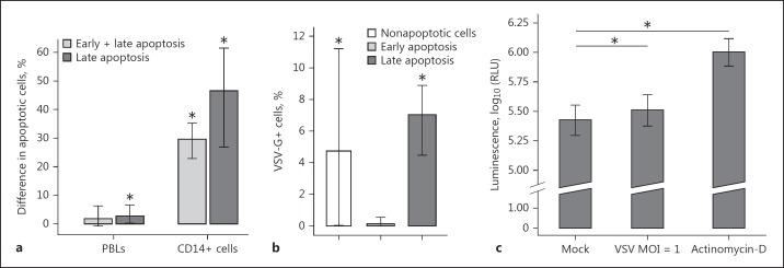
Fig. 7.
VSV infection caused apoptosis in PBLs and CD14+ cells. Isolated PBLs were mock- or VSV-infected or incubated with actinomycin for 18 h. a Analysis was applied to PBLs within the combined lymphocyte-monocyte gate or to CD14+ cells only (n = 7). The bars with whiskers represent the average value with its 95% confidence interval obtained by a t test on data transformed using the Box-Cox method, * p < 0.05. b The percentage of VSV-infected (VSV-G+) cells within cells in different stages of apoptosis (n = 7). The average value with its 95% confidence interval is presented (t test on data transformed using the Box-Cox method, * p < 0.05). c Caspase-3/7 activity (n = 10). The average value with a 95% confidence interval is shown (one-sided exact permutation test for paired data, * p < 0.05).

