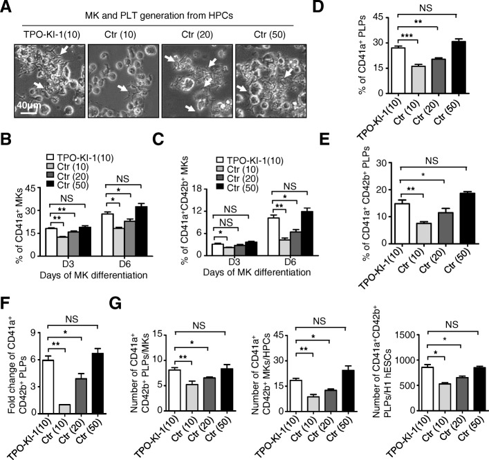
Fig. 5.
TPO knock-in partially replaces extrinsic TPO. a Representative morphology of proplatelets (white arrows) among indicated groups at day 6 (D6) during MK differentiation. Scale bar = 40 μm. b, c Flow cytometry analysis showing percentage of CD41a+ (b) or CD41a+CD42b+ (c) MKs in indicated groups at day 3 (D3) and D6, respectively. Data shown as mean ± SEM (n = 3). *P < 0.05; **P < 0.01; NS, not significant. d, e Flow cytometer analysis for percentage of CD41a+ (d) or CD41a+ CD42b+ (e) platelets in indicated groups, respectively. Data shown as mean ± SEM (n = 3). *P < 0.05; **P < 0.01; NS, not significant. f Fold change of CD41a+CD42b+ PLPs in indicated groups. Data shown as mean ± SEM (n = 3). *P < 0.05; **P < 0.01; NS, not significant. g Comparative analyses of CD41a+CD42b+ PLTs per MKs or seeded H1 hESCs, MKs per HPCs, at day 6 of megakaryocytic differentiation among four groups. Data shown as mean ± SEM (n = 3). *P < 0.05; **P < 0.01; NS, not significant. Ctr control, hESC human embryonic stem cell, HPC hematopoietic progenitor cell, MK megakaryocyte, PLT platelet, TPO thrombopoietin, PLP platelet-like particles

