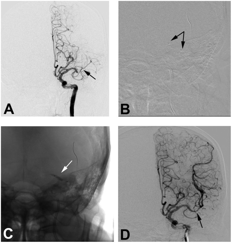
Figure 2.
An 81-year-old male patient with hypertension and coronary heart disease for 10 years presented with paralysis of the right upper limb for six hours. (a) Digital subtraction angiography confirms occlusion of the left middle cerebral artery (MCA) M1 segment (black arrow). (b) A 2 × 15 mm balloon (Gateway) is deployed at the occluded segment (black arrows). (c) The balloon inflates at the occluded segment (white arrow). (d) After three rounds of inflation, final angiography shows good perfusion with 10% fixed focal stenosis at the joint site between the MCA M1 and M2 segment with good distal perfusion (black arrow).

