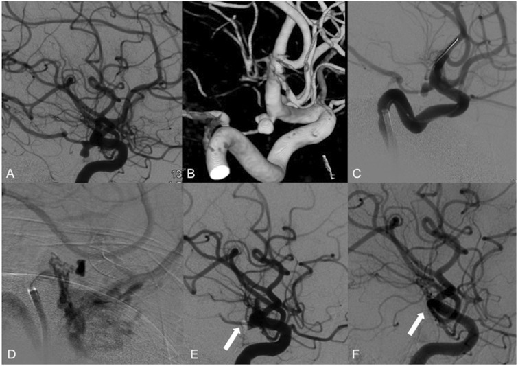
Figure 2.
Use of the PED in an incidentally found PComA aneurysm. (a) Selective right ICA contrast injection shows an incidental 6 × 4 × 3 mm PComA aneurysm. (b) Acquisition of cone-beam CT 3D reconstruction images for procedure planning and better visualization of the vessel anatomy. (c) A 3.75 × 18 mm PED was placed across the right PcomA. (d) Flow stagnation is immediately seen within the aneurysm lumen after placement of the device. ((e), arrow) There is also decreased flow across the right PComA post-device placement upon completion of the procedure. ((f), arrow) Six-month follow-up angiography shows complete occlusion of the aneurysm and PcomA. PED: Pipeline embolization device; PComA: posterior communicating artery; ICA: internal carotid artery; CT: computed tomography; 3D: three-dimensional.

