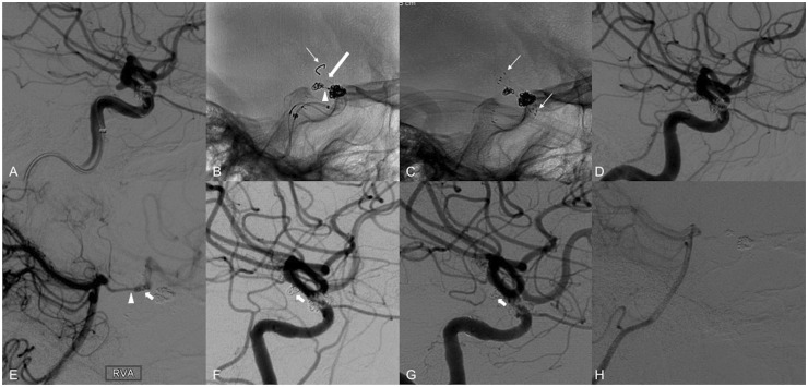
Figure 3.
Use of the PED in conjunction with a self-expanding laser-cut stent. (a) Right ICA angiography reveals a 2.5 × 2.9 × 2.2 mm PComA aneurysm with the branch arising from the dome of the aneurysm. The patient underwent previous stent-assisted coiling of a right paraophthalmic aneurysm. (b) A 3 mm × 4 cm coil was placed within the aneurysm lumen via a microcatheter (arrow head) which was jailed by the 3.0 × 14 mm Pipeline Flex device (thin arrow on tip of delivery wire). Unfortunately, the PED foreshortened and covered only the proximal portion of the aneurysm neck (thick arrow). ((c), arrows on distal and proximal markers of the stent) A 3 × 15 mm Neuroform EZ stent (Stryker Neurovascular, Fremont, CA) was then telescopically placed across the distal end of the PED, successfully covering the aneurysm lumen and anchoring the PED against the vessel wall. (d) Follow-up angiogram demonstrating patency of the PComA. (e) Right vertebral artery angiogram post-treatment also demonstrates patency of the PComA (arrow head) and flow through the aneurysm (arrow) into the anterior circulation. (f) Six-month follow-up angiography demonstrates complete occlusion of the PComA and aneurysms. (g) One-year follow-up angiography shows unchanged complete occlusion of the PComA and aneurysm. (h) Similar to at six months (image not shown), the right vertebral artery angiogram at one year reveals no flow through the PComA. PED: Pipeline embolization device; PComA: posterior communicating artery; ICA: internal carotid artery.

