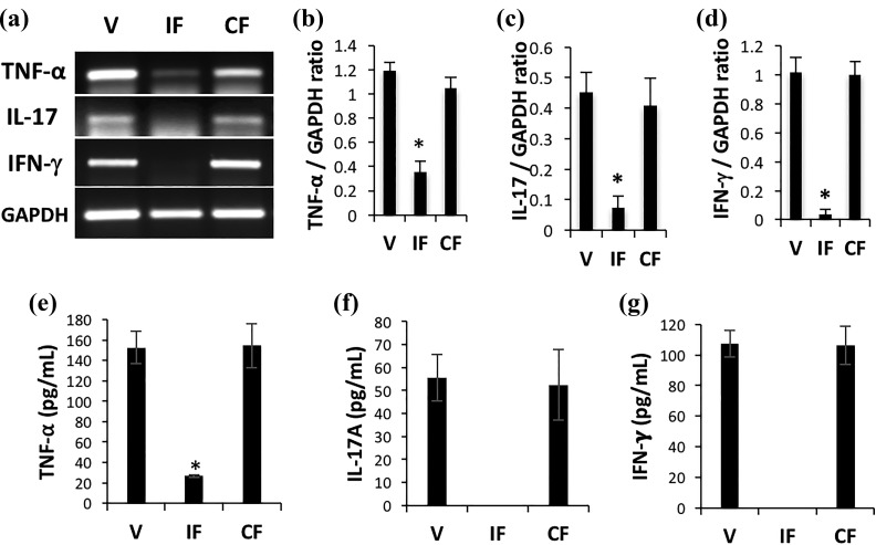
Figure 3.
IDO fibroblast therapy resulted in decreased expression of proinflammatory cytokines in skin. IDO-expressing fibroblasts, control fibroblasts, or vehicle solution were injected intraperitoneally into C3H/HeJ mice at the time of AA induction. Total RNA was extracted from skin samples 20 weeks post-AA induction and was analyzed using reverse transcriptase polymerase chain reaction (RT-PCR). Panel (a) shows representative RT-PCR results for TNF-α, IL-17, and IFN-γ in vehicle (V), IDO fibroblast (IF), and control fibroblast (CF) groups. Glyceraldehyde 3-phosphate dehydrogenase (GAPDH) was run as loading control. Panels (b) to (d), respectively, show average relative cytokine mRNA expression ± SD of TNF-α, IL-17, and IFN-γ in skin of different treatment groups. Panels (e) to (g) show quantitative measurement of these inflammatory cytokines at protein level in skin of different treatment groups using a cytometric bead assay. * denotes statistically significant difference between IDO and the two other control groups (n = 5, P < 0.001).

