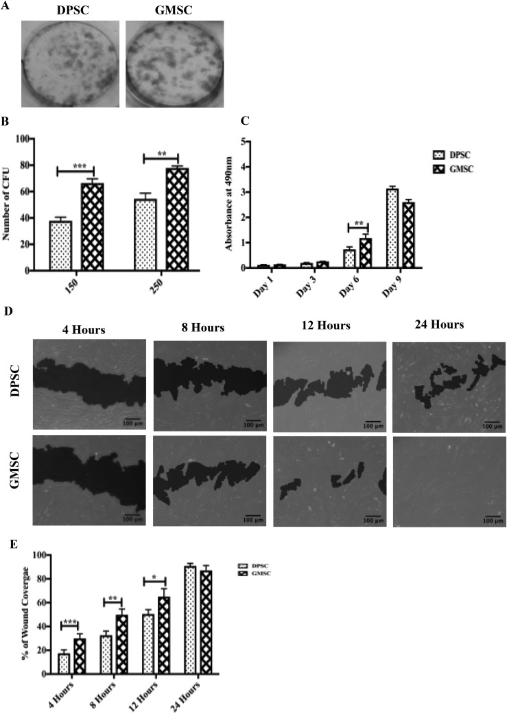
Fig. 1.
Gingival mesenchymal stem cells (GMSCs) and dental pulp stem cells (DPSCs) showed different clonogenic and proliferation potentials. (A) Representative images of colony-forming units (CFUs) stained with crystal violet after 20 d in culture. (B) An increase in the formation of CFUs was observed for both concentrations (150 cells and 250 cells) for GMSCs compared to DPSCs with a P < 0.05. (C) Quantification of cell proliferation between DPSCs and GMSCs incubated at different time points (1, 3, 6, and 9 d). An increase in the proliferation of GMSCs compared to DPSCs was observed between day 6 compared to DPSCs with a P < 0.05. (D) In vitro migration comparison between DPSCs and GMSCs based on a 24-h scratch wound healing assay. (E) GMSCs display a better migratory capacity compared to DPSCs for 4, 8, and 12 h (P < 0.05). At 24 h, no significant change in the proliferation was observed. All data are represented as a mean with the associated standard error of the mean (n = 3) of a minimal 3 donors.

