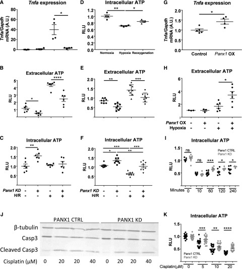
Figure 4.
ATP release and/or intracellular retention in kidney tubule epithelial cells are key factors in protection from injury. (A–F) Pannexin1 (Panx1)-deficient (Panx1 knockdown [KD], +) and scrambled control (Panx1 KD, −; referred to as Panx1 control in Results) murine proximal tubule cell line (TKPTS) clones were created using the CRISPR/Cas9 system. Hypoxia-reoxygenation (H/R) injury was simulated in vitro by exposing cells to (A–C, G, and H) 60 minutes or (D–F) 360 minutes of hypoxia under mineral oil (or culture medium for sham conditions) followed by a period of reoxygenation in culture medium. Extracellular and intracellular ATP content was measured after a (B and C) 15-minute or (D–F) 60-minute reoxygenation period, and it is expressed as fold change compared with sham control cells (relative light units [RLUs]). (C) Tnfa mRNA levels were measured in cell lysates after 60 minutes of reoxygenation as an indicator of H/R-induced injury. (D) Intracellular ATP during normoxia, after 360 minutes of hypoxia alone, and after 360 minutes of hypoxia followed by 60 minutes of reoxygenation. (G and H) Transient overexpression of Panx1 causes cells to release more ATP in stress conditions. TKPTS cells were transfected using lipofectamine (mock transfection, control; −) or lipofectamine and plasmid to observe Panx1 overexpression (OX; +). (G) Tnfa mRNA levels were measured in cell lysates after 60 minutes of reoxygenation as an indicator of H/R-induced injury. (H) Extracellular ATP content was measured after a 15-minute reoxygenation period, and it is expressed as fold change compared with sham control cells. (I) Intracellular ATP decreased after addition of antimycin A but recovered more rapidly in Panx1-deficient cells. Antimycin A (20 μm) was added to cells, and intracellular ATP was measured 0, 10, 60, 120, or 240 minutes later. (J) Western blot of KD and control TKPTS cells treated with 20 or 40 μM cisplatin for 16 hours showing expression of caspase3 and the amount of cleaved caspase3, with β-tubulin used as loading control. Quantification is in Supplemental Figure 4C. (K) Decrease in intracellular ATP after incubation with cisplatin is smaller in Panx1-deficient cells. Treatment of cells with 5–20 μM cisplatin for 18 hours. Values are shown as fold change in RLU, with each group compared with their respective nontreated control. CTRL, control; Gapdh, glyceraldehyde-3-phosphate dehydrogenase. *P<0.05; **P<0.01; ***P<0.001; ****P<0.001.

