Figure 1.
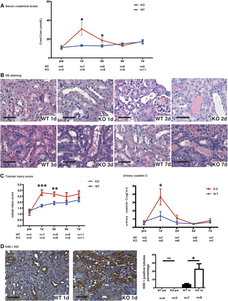
Caspase-3 deficiency aggravates IRI-induced tubular injury. (A) Serum creatinine levels in wild-type (WT) and caspase-3−/− (KO) mice at baseline (pre-op), and 1, 2, 3, and 7 days post-IRI. (B) Representative hematoxylin and eosin (H&E)–stained renal sections from WT and KO mice at 1, 2, 3, and 7 days post-IRI (original magnification ×200). (C) Left panel: mean tubular injury scores of ten randomly chosen high-power fields (original magnification ×200) in mice kidney sections post-IRI. Right panel: urinary levels of cystatin C in WT and KO mice at baseline (pre-op) and 1, 2, 3, or 7 days post-IRI. (D) Left panels: representative Kidney Injury Molecule 1 (KIM-1) immunohistochemistry in renal cortical sections from WT and KO mice at day 1 post-IRI (magnification 200×). Right panel: quantification of KIM-1 immunohistochemistry-stained murine renal cortical sections at baseline (pre-op) and 1 day post-IRI. All scale bars, 50 μm. Values are mean±SEM. *P<0.05, compared between WT and KO at the same time point.

