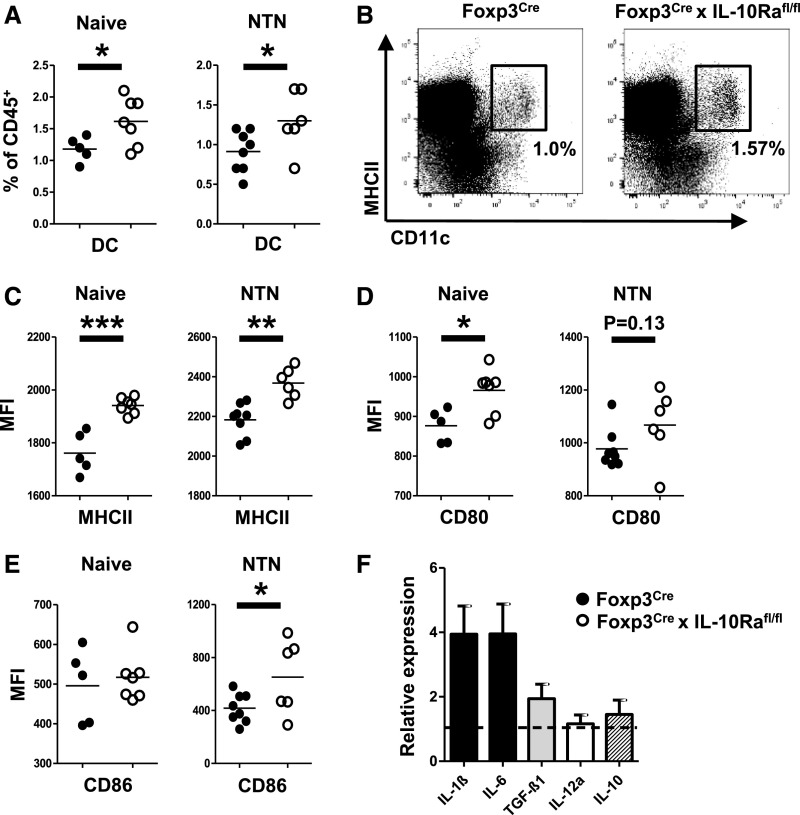
Figure 4.
Absence of IL-10 receptor a (IL-10Ra) on regulatory T cells results in a Th17-promoting dendritic cell (DC) phenotype. (A) FACS analysis of CD11c+MHCII+ DCs in spleens of naïve mice and at 3.5 days after induction of NTN. (B) Representative FACS plot showing DCs in spleens of naïve mice. (C) FACS analyses of MHCII mean fluorescence intensity (MFI) on splenic DCs of naïve mice and at 5.5 days after induction of NTN. FACS analyses of (D) CD80 MFIs and (E) CD86 MFIs on splenic DCs of naïve mice and at 3.5 days after induction of NTN. Group sizes in A–E were n=5 versus n=7 naïve mice and n=8 versus n=6 nephritic mice. (F) Quantitative RT-PCR analyses of indicated cytokines from splenic DCs isolated at 36 hours after antigen challenge (n=5 mice). The dotted line represents DCs from wild-type mice (n=5 mice). Bar graphs show means, and error bars indicate SEM. Circles represent individual animals, and horizontal lines show mean values. *P<0.05; **P<0.01; ***P<0.001.

