Figure 8.
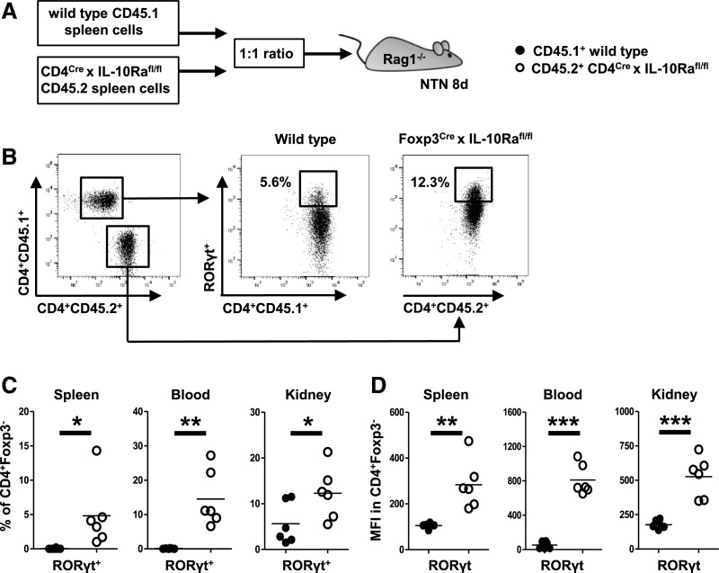
IL-10 receptor a (IL-10Ra) signaling directly controls Th17 cells but not regulatory T cell development and fitness. (A) Overview of the experimental setup. Spleen cells from CD45.1+ mice with IL10Ra-sufficient CD4+ T cells and CD45.2+ mice with IL10Ra-deficient CD4+ T cells were cotransferred into n=6 Rag1−/− recipient mice, and NTN was induced. (B) Representative FACS plot of CD45.1 and CD45.2 CD4+ T cells in the kidney of Rag1−/− recipients at day 8 of NTN (left panel). Representative FACS plots indicating percentages of RORγt+ Th17 cells among both CD45 subtypes (center and right panels). (C) Quantification of RORγt+ Th17 cells among both CD45 subtypes in the indicated compartments. (D) Mean fluorescence intensity (MFI) of RORγt protein in CD4+ Teff in indicated compartments. Circles represent individual animals, and horizontal lines show mean values. *P<0.05; **P<0.01; ***P<0.001.

