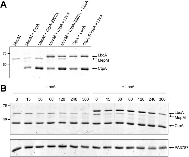
FIG 7 .
In vitro proteolysis of MepM. (A) CtpA degrades MepM and is enhanced by LbcA. 2 μM concentrations of the indicated C-terminal His6-tagged proteins was incubated for 3 h at 37°C. Samples were separated on a 12.5% SDS-PAGE gel, which was stained with Coomassie brilliant blue. Approximate positions of molecular mass marker proteins (in kilodaltons) are indicated on the left-hand side. (B) Time course. Reaction mixtures contained 2 µM CtpA-His6 and MepM-His6 either without (−LbcA) or with (+LbcA) 2 µM LbcA-His6. Reactions were terminated at the indicated time points (minutes) by adding SDS-PAGE sample buffer and boiling. As a negative control, the same experiment was done using PA3787 in place of MepM. Samples were separated on 12.5% SDS-PAGE gels, which were stained with Coomassie brilliant blue. Approximate positions of molecular mass marker proteins (in kilodaltons) are indicated on the left-hand side. PA3787 ran at the midway point between marker proteins of 37 and 25 kDa, which were above and below, respectively, the region of the gel shown in the figure.

