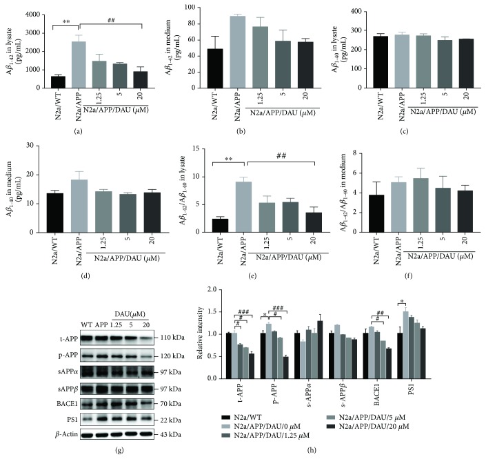
Figure 2.
DAU inhibited APP processing and Aβ accumulation. Levels of Aβ 1–42 (a, b), Aβ 1–40 (c, d), and Aβ 1–42/Aβ 1–40 (e, f) of cell lysates (a, c, e) and cell culture media (b, d, f) as a function of DAU concentration were determined by ELISA. Levels of t-APP, p-APP, s-APPα, s-APPβ, BACE1, and PS1 were determined by Western blot analysis (g, h). β-Actin was used as a loading control. N = 3. Data show the mean ± SEM. ∗ P < 0.05 and ∗∗ P < 0.01 compared to N2a/WT cells. # P < 0.05, ## P < 0.01, and ### P < 0.001 compared to untreated N2a/APP cells.

