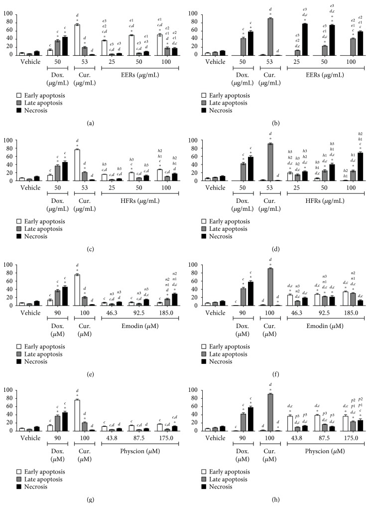
Figure 3.
Cell death analysis in HSC-3 cells. Cytomorphological viability assay for (a) EERs, (c) HFRs, (e) emodin, and (g) physcion. Annexin-V assay for (b) EERs, (d) HFRs, (f) emodin, and (h) physcion. Comparison with significant statistical differences between treatments: vehicle (∗); doxorubicin (d); curcumin (c); EERs: 25 μg/mL (e1); EERs: 50 μg/mL (e2); EERs: 100 μg/mL (e3); HFRs: 25 μg/mL (h1); HFRs: 50 μg/mL (h2); HFRs: 100 μg/mL (h3); emodin: 46.3 μM (n1); emodin: 92.8 μM (n2); emodin: 185.0 μM (n3); physcion: 43.8 μM (p1); physcion: 87.5 μM (p2); physcion: 175.0 μM (p3); and cell status: early apoptosis, late apoptosis, and necrosis. Results are expressed as mean of three independent experiments ± standard error (M ± SE), analyzed by one-way ANOVA with Tukey's posttest, p ≤ 0.05.

