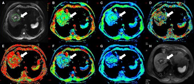
Figure 5.

Seventy‐two‐year‐old female with liver metastases(arrow). (A) is b value of 50 s/mm2 of DWI, and (B‐G) are pseudocolor of ADC, D t, D p, f p, DDC and α. The values of lesion were 0.923 × 10⁻³, 0.807 × 10⁻³, 1.44 × 10⁻² mm2/s, 0.345, 0.608 × 10⁻³ mm2/s, and 0.702, respectively. (H) is T2 image
