Figure 5.

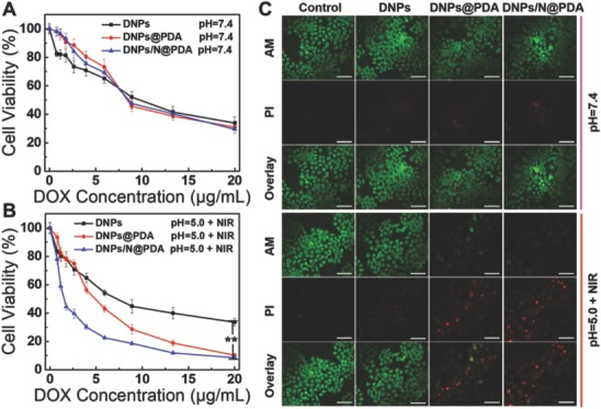
A,B) In vitro cytotoxicity of DNPs, DNPs@PDA, and DNPs/N@PDA at pH 7.4 and pH 5.0 with NIR laser irradiation (808 nm) of 5 W cm−2 for 1 min at different DOX concentrations on HeLa cells after 48 h incubation (**p < 0.01 versus DNPs/N@PDA group with NIR irradiation). C) Confocal fluorescence microscopy images of HeLa cells of different treatments for 48 h costained with Calcein‐AM (green, live cells) and propidium iodide (PI) (red, dead cells) before and after laser illumination (808 nm, 5 W cm−2, 1 min). The DOX concentration was fixed at 5 µg mL−1. Scale bars: 100 µm.
