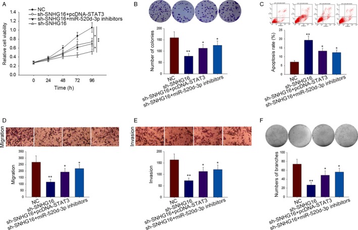
Figure 5.

The effects of SNHG6‐miR‐520d‐3p‐STAT3 pathway on HemECs cell activities. A‐B, The proliferation ability of indicated HemECs was detected by MTT and colony formation assays. C, The apoptosis of transfected HemECs cell was detected by flow cytometry analysis. D‐E, The migration and invasion of HemECs cells were detected using transwell assay. F, Tube formation assay was carried out in indicated HemECs cells to investigate the vasoformation. Data were acquired from multiple experiments for mean ± SD. *P < .05, **P < .01 compared with controls
