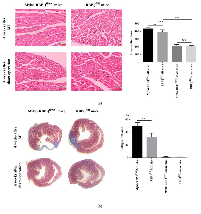
Figure 2.
Higher collagen density in the infarct area in the Myh6-RBP-J fl/wt mice following MI. (a) 25 visual fields at the border zone were randomly selected from 4 mice for each group, and the cross-sectional area of cardiomyocytes was estimated (n = 4, ∗∗P < 0.01 and ∗∗∗P < 0.001). Data are shown as means ± SD. (b) 4 weeks after operation, the hearts of Myh6-RBP-Jfl/wt-MI mice, RBP-Jfl/fl-MI mice, Myh6-RBP-Jfl/wt-sham mice, and RBP-Jfl/fl-sham mice were sectioned and stained using Masson staining. Cardiac fibrosis was assessed by calculating the ratio of fibrotic scar (blue) average circumferences to left ventricular average inner circumferences (n = 4, ∗∗P < 0.01). Data are shown as means ± SD.

