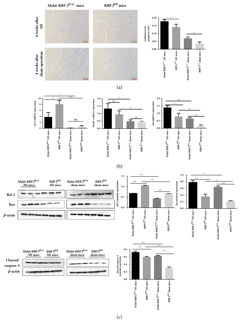
Figure 3.
RBP-J knockout increased cardiomyocyte apoptosis following MI. (a) TUNEL staining showed that the number of apoptotic cells was increased in the border zone of ischemic heart tissue of Myh6-RBP-Jfl/wt mice compared with RBP-Jfl/fl mice with MI (original magnification: × 200, brown indicates TUNEL-positive nuclei and blue indicates nonapoptotic cells). The apoptosis rates of tissues in each group (Myh6-RBP-Jfl/wt-MI mice, RBP-Jfl/fl-MI mice and Myh6-RBP-Jfl/wt-sham mice, and RBP-Jfl/fl-sham mice) were detected (n = 6, ∗P < 0.05). Each value represents the mean ± SD. (b) Expression of bcl-2 and bax mRNA of different groups ((Myh6-RBP-Jfl/wt-MI mice, RBP-Jfl/fl-MI mice and Myh6-RBP-Jfl/wt-sham mice, and RBP-Jfl/fl-sham mice). (c) The protein expression level of bcl-2, bax, cleaved-caspase 3 by western blot analysis. The results are represented as mean ± SD (n = 6, ∗P < 0.05, ∗∗P < 0.01).

