Fig. 1.
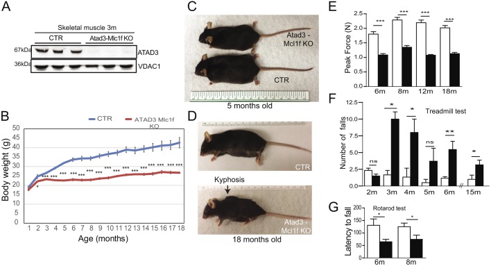
Creation of muscle-specific Atad3 KO mice. (A) Western blot analysis of ATAD3 protein levels in mitochondria from hind-limb skeletal muscle of 3-month-old animals. VDAC1 was used as a loading control. (B) Body weight measurement: Atad3-Mlc1f male KO mice (red) showed a significantly reduced weight compared with age-matched control (CTR) mice (blue) from 2 months of age onwards (n=12 in KO group and n=15 in control group). (C,D) Representative images of control and Atad3-Mlc1f KO male mice of 5 (C) and 18 (D) months of age, showing decreased body weight and development of kyphosis in the Atad3-Mlc1f KO male mice compared with the controls. (E) Atad3-Mlc1f KO mice showed reduced muscle strength compared with control mice at 6 months of age (n=10). (F,G) Treadmill (F, n=8) and rotarod tests (G, n=12) performed by Atad3-Mlc1f KO and control mice. Data are mean±s.e.m. P-values were calculated by Student's t-test. n.s., nonsignificant; *P<0.05, **P<0.01 and ***P<0.0001.

