Fig. 4.
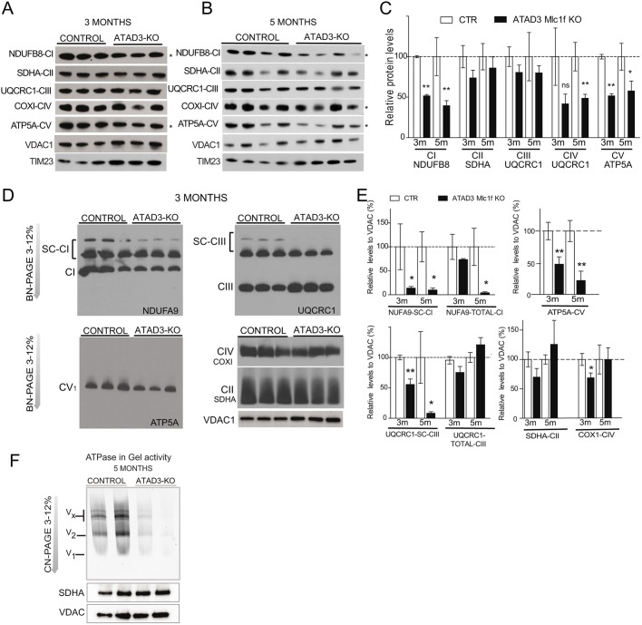
Analysis of OXPHOS complexes in control and Atad3-Mlc1f KO skeletal muscle mitochondria. (A,B) Western blot analysis of mitochondrial protein levels in purified mitochondria from the muscle of 3- and 5-month-old control and Atad3-Mlc1f KO mice. (C) Quantification of the steady-state levels of respiratory subunits shown in A and B. VDAC1 was used as a loading control. (D) BN-PAGE analysis of respiratory chain supercomplexes followed by western blot analysis using antibodies against complex I (anti-NDUFA9), complex II (anti-SDHA), complex III (anti-UQCRC1), complex IV (anti-COXI), and complex V (anti-ATP5Α) in mitochondrial extract from muscles of 3-month-old control and Atad3-Mlc1f KO mice. VDAC1 was used as a loading control. (E) Quantification of relative levels of OXPHOS complexes and supercomplexes. (E) In-gel activity of complex V (ATP synthase) in muscle mitochondria from 5-month-old control and Atad3-Mlc1f KO mice. Complex V activity in gel is reduced in the mitochondrial extract from Atad3-Mlc1f KO muscle. Data are mean±s.e.m. P-values were calculated by Student's t-test. ns, nonsignificant; *P<0.05 and **P<0.01.

