Fig. 6.
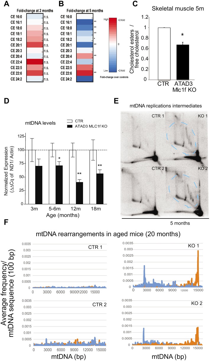
Loss of ATAD3 in muscle impairs cholesterol trafficking and results in mtDNA depletion, replication stalling and rearrangements. (A,B) Lipidomic analysis of the different cholesterol esters species related to total cholesterol in gastrocnemius muscle of control and Atad3-Mlc1f KO mice aged 2 (A) and 5 (B) months. Data are expressed as fold change over the controls and represented as heat maps. (C) Quantification of the total cholesterol esters/free cholesterol in muscle from 5-month-old control and Atad3-Mlc1f KO mice. Data are mean±s.e.m. n=4 in control group; n=5 in KO group. P-values were calculated by Student's t-test. (D) Analysis of mtDNA levels in skeletal muscle from Atad3-Mlc1f KO and control mice of 3, 5, 12 and 18 months age (tibialis anterior). mtDNA/nDNA levels were measured by real-time PCR from four control and five KO animals. Data are mean±s.e.m. P-values were calculated by Student's t-test. (E) Analysis of mtDNA replication intermediates (bands within the dashed blue lines) in the skeletal muscle of Atad3-Mlc1f KO and control mice. Mitochondrial nucleic acids from two Atad3-Mlc1f KO and two control quadriceps were digested with ClaI and separated by 2D-AGE; the blot was hybridized with a part of the cytochrome B sequence of the murine mitochondrial genome. (F) Frequencies of breakpoint positions in muscle from 20-month-old Atad3-Mlc1f KO and control mice obtained by next generation sequencing (NGS). Breakpoint position of 5′ bases shown in blue and 3′ bases in orange as 100 bp rolling average. The numbering corresponds to the murine mtDNA reference sequence (NC_005089). n.s., nonsignificant; * P<0.05 and **P<0.01.

