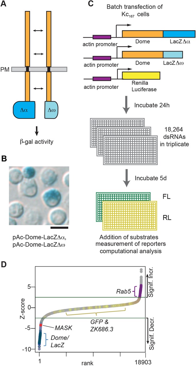
Fig. 1.

A split β-galactosidase genome-wide RNAi screen to identify modulators of Dome dimerisation and levels. (A) Schematic representation of the Dome–β-galΔα and Dome–β-galΔω complementation assay. PM, plasma membrane. (B) Drosophila Kc167 cells transiently transfected with plasmids expressing the proteins shown in A show β-galactosidase activity as determined by X-gal staining. Scale bar: 10 μm. (C) Workflow of the genome-wide RNAi screen for modulators of Dome dimerisation and levels as undertaken in Drosophila Kc167 cells. FL, firefly luciferase; RL, Renilla luciferase. (D) Ranked Z-scores from the genome-wide RNAi screen. Green lines illustrate Z-score cut-offs of significant increase or significant decrease. Controls are shown (Dome, LacZ, GFP, ZK686.3, Rab5) with MASK highlighted in red.
