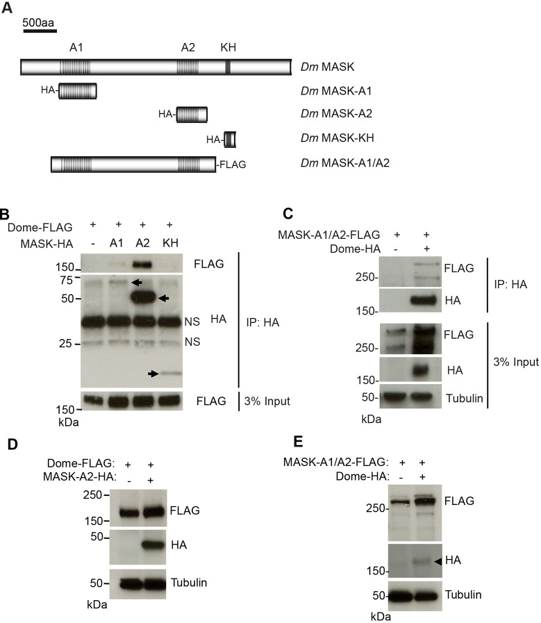
Fig. 3.
MASK physically associates with Dome. (A) Schematic representation of Drosophila MASK protein and constructs used in this study. (B) Immunoprecipitation (IP) of the indicated HA–MASK constructs from Kc167 cells also expressing Dome–FLAG (arrows). Dome–FLAG is co-immunoprecipitated with HA–MASK-A1 and HA–MASK-A2. Levels of Dome–FLAG present in the input lysate are shown in the lower panel. NS, non-specific band. (C) Co-precipitation of MASK-A1/A2-FLAG following immunoprecipitation of Dome–HA. Levels of MASK-A1/A2-FLAG, Dome-HA and α-Tubulin present in the total Kc167 cell lysates are shown in the lower panels. (D) Steady-state levels of Dome–FLAG expressed in Drosophila Kc167 cells are increased following the co-expression of HA–MASK-A2. Levels of α-Tubulin indicate loading parity. (E) Steady-state levels of MASK-A1/A2–FLAG expressed in Drosophila Kc167 cells are increased following the co-expression of Dome–HA (arrowhead). Levels of α-Tubulin indicate loading parity.

