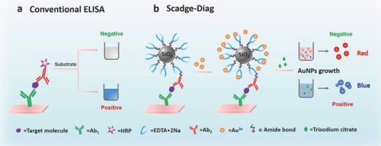
Figure 1.

Schematic representation of conventional sandwich ELISA format and Scadge‐Diag bioassay performed in 96‐well polystyrene (PS) plates. a) In the conventional colorimetric ELISA, enzymatic biocatalysis dominates the color change of substrate. b) In the Scadge‐Diag bioassay, leveraging on the metal sequestration of a chelating agent (EDTA•2Na) for dominating gold nanoparticle generation (Scadge), EDTA•2Na substituting enzyme is employed for bioassay and this detection is enhanced through a silica nanocarrier as a signal amplifier.
