
Fig. 2.
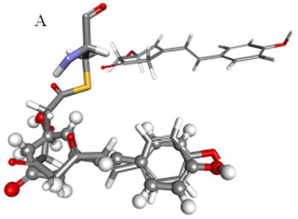
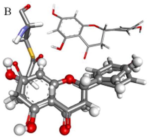
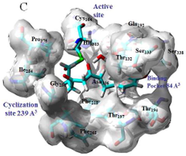
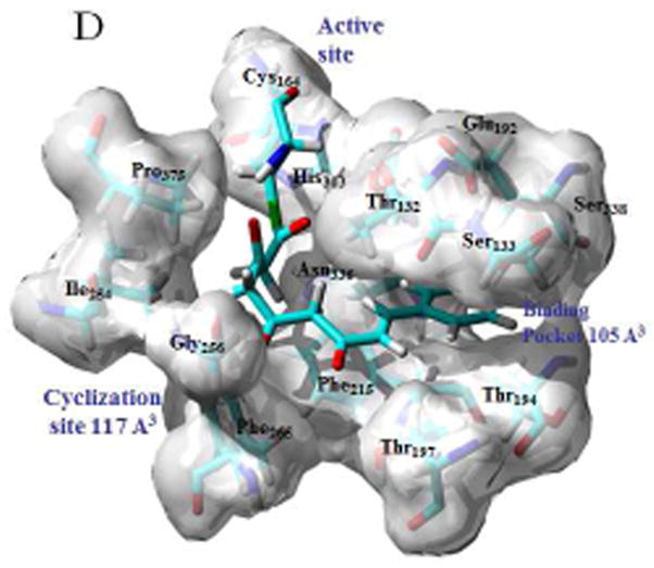
Modeled structures for the bound tetraketide in STS (A) and CHS (B) are shown overlaid with the bound resveratrol and naringenin ligands, with the crystal structures for the ligands shown in inset; binding pockets, active sites and cyclization sites in STS (C) and CHS (C), showing the calculated cavity volumes for the cyclization sites and binding pockets.