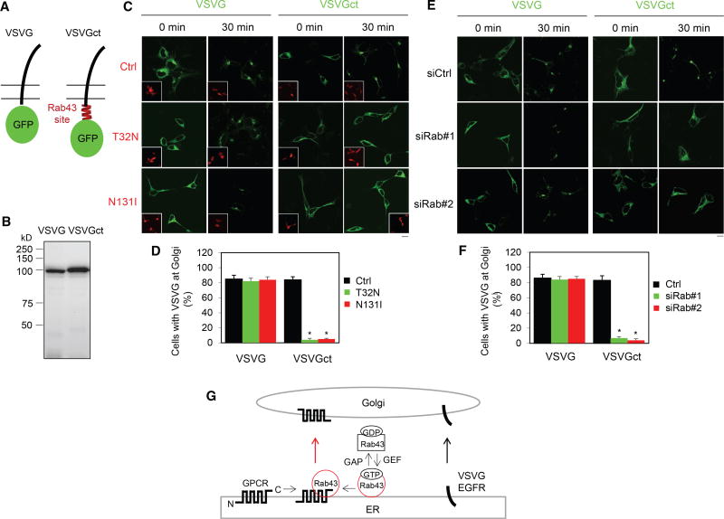
Figure 7. Effect of the Rab43-Binding Domain Identified in the AT1R CT on the ER-Golgi Transport of VSVG.
(A) A diagram showing the generation of the VSVG chimera containing the Rab43-binding domain identified in the AT1R CT (VSVGct).
(B) Expression of VSVG and VSVGct in HEK293 cells by immunoblotting using GFP antibodies.
(C) Effect of Rab43 mutants on the ER-to-Golgi transport of VSVG and VSVGct. HEK293 cells were transfected with VSVG-GFP or VSVGct-GFP together with dsRed-C1 or dsRed-Rab43 mutants. The cells were cultured at 40°C for 24 hr (0 min) and then shifted to 32°C for 30 min.
(D) Quantitation of the data shown in (C).
(E) The effect of Rab43 siRNA on the ER-to-Golgi transport of VSVG and VSVGct.
(F) Quantitation of the data shown in (E).
(G) A model depicting the roles of Rab43 in the ER-to-Golgi transport of GPCRs as well as their sorting at the ER after synthesis by virtue of its ability to interact with the receptors in an activation-dependent manner (see text for details).
The data shown in (D) and (F) are percentage of cells with VSVG expression at the Golgi, with a total of 100 cells counted in each experiment, and are presented as the mean ± SE (n = 3–5). Scale bars, 10 µm. *p < 0.05 versus Ctrl.

