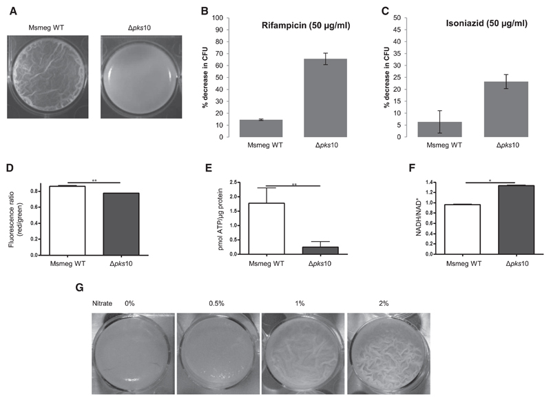
Figure 4. Effect of pks10 Gene Knockout on the Mycobacterial Biofilm.
(A) Biofilm formed by the WT and Δpks10 strains.
(B) Percent decrease in colony-forming units (CFUs) upon treatment of Msmeg biofilm with rifampicin.
(C) Percent decrease in CFUs upon treatment of Msmeg biofilm with isoniazid.
(D) Assessment of the membrane potential of cells in biofilm cultures of the WT and Δpks10 strain based on the potential dependent internalization of the carbocyanine dye 3,3′-diethyloxacarbocyanine iodide.
(E) Level of intracellular ATP in biofilm cultures of the WT and Δpks10 strain.
(F) Estimation of intracellular NADH/NAD+ levels in biofilm cultures of the WT and Δpks10 strains.
(G) Effect of exogenous addition of nitrate on the biofilm of the Msmeg Δpks10 strain.
Statistical significance was analyzed by two-tailed Student’s t test. *p < 0.05, **p < 0.01. All error bars represent ± SEM. See also Figure S7.

