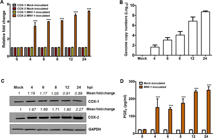
Fig 2. COX-2 mRNA and protein levels increase upon murine norovirus (MNV) infection with subsequent production of PGE2.
(A and B) Cultured RAW264.7 cells were mock-infected or infected with MNV (MOI, 1 TCID50/ml) for the indicated time points, and the expression of COX-1 and COX-2 mRNAs, and MNV viral RNA were determined by quantitative real time PCR. The mRNA expression levels of COX-1 and COX-2 were normalized to β-actin mRNA and are presented as a fold induction as compared with the mock-infected cells. (C) Monolayers of RAW264.7 cells were infected with or without MNV (MOI, 1 TCID50/ml) for the indicated time points, and the levels of the COX-1, COX-2, and GAPDH proteins were analyzed by Western blot analysis. GAPDH was used as a loading control. The intensity of each target protein relative to GAPDH was determined by densitometric analysis and is indicated above each lane. (D) Cell culture supernatants harvested from the cells infected with or without MNV at the indicated time points were checked for the presence of PGE2 by ELISA. The levels of PGE2 in the supernatants were compared between mock-and MNV-infected groups. The presented data are depicted as means and standard errors of the mean from three different experiments. Statistical analyses were performed using one-way analysis of variance. ***p < 0.0001.

