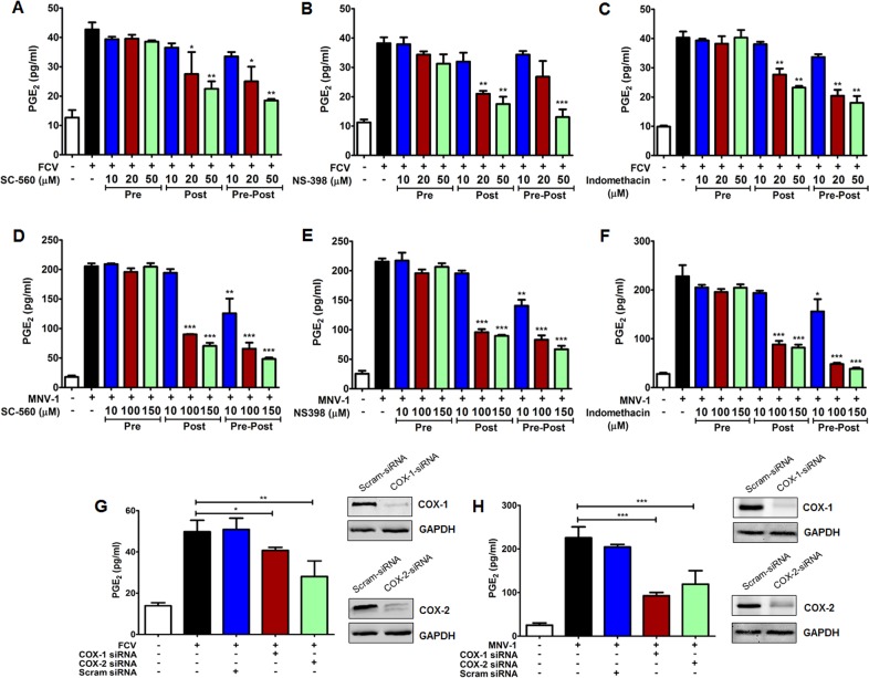
Fig 3. Influence of COX inhibitors on PGE2 production during feline calicivirus (FCV) and murine norovirus (MNV) replication.
(A–F) CRFK and RAW264.7 cells were treated with a selective COX-1 inhibitor (SC-560), a selective COX-2 inhibitor (NS398), or a nonselective COX inhibitor (indomethacin) at the indicated time points. Cells pretreated with each inhibitor were washed to remove each inhibitor and then infected with FCV (MOI, 1 FFU/ml) or MNV (MOI, 1 TCID50/ml) strains (Pre). After virus adsorption, the inhibitor(s) were added in the maintenance media (Post), or before virus inoculation and maintained throughout the course of the infection (Pre-Post). The levels of PGE2 in the supernatants harvested at 4 hpi for FCV and 24 hpi for MNV were determined by ELISA. The PGE2 levels from virus-infected supernatants were compared between the mock- and drug-treated groups. (G and H) Confluent CRFK cells (G) and RAW264.7cells (H) were transfected with COX-1 and COX-2, or scrambled (Scram) siRNAs, and then infected with FCV (MOI, 1 FFU/cell) for 4 h or MNV (MOI, 1 TCID50/ml) for 24 h. Then, supernatants were collected and PGE2 levels were detected by ELISA. (Insets) CRFK cells (G) and RAW264.7 cells (H) transfected with COX-1, COX-2, or scrambled siRNAs were harvested and subjected to Western blot analyses. GAPDH was used as a loading control. The results are presented as means and standard errors of the mean of three independent experiments. Statistically analyses were performed using one-way analysis of variance. *p < 0.05; **p < 0.001; ***p < 0.0001.

