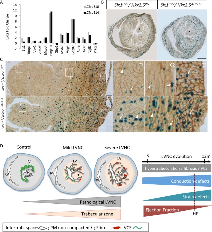
Fig 9. Molecular markers associated with hypertrabeculated Nkx2-5 mutant hearts and mouse models of pathological LVNC.
(A) Quantitative real-time PCR performed for a list of selected genes. The housekeeping gene used was RPL32. (B-C) X-gal (B) and Pecam-1 (C) co-immunostaining on transversal sections of the mid-ventricle from Six1LacZ/+::Nkx2-5+/+ and Six1LacZ/+::Nkx2-5ΔTrbE10 adult hearts. Dotted lines indicate the boundary between the trabecular and compact zone of the left ventricular myocardium; asterisks, the papillary muscles (PM); arrows, β-positive cardiomyocytes; arrowheads, β-gal-positive endothelial cells. (C1-C3) High magnifications of boxes at the level of the PM (C1), the trabecular/compact boundary (2) and the epicardium (3) on the LV. Scale bar B = 1mm; C = 100μm; C1-3 = 50μm. (D) Mouse models of mild and severe hypertrabeculation associated with pathological features including intertrabecular recesses, non-compacted papillary muscles, fibrosis and VCS hypoplasia. The degree of hypertrabeculation is relative to the extent of trabecular zone affected. The follow-up of cardiac function shows a correlation of impaired Ejection Fraction with conduction and strain defects in absence of aggravation of the hypertrabeculation phenotype with age. HF: Heart failure.

