Figure 1.

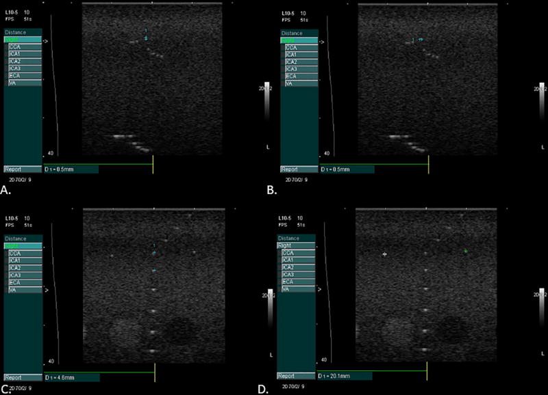
Phantom images taken for quality assurance to measure axial (panel A) and lateral (panel B) resolution and to confirm calibration in the vertical (panel C) and horizontal planes (panel D).

Phantom images taken for quality assurance to measure axial (panel A) and lateral (panel B) resolution and to confirm calibration in the vertical (panel C) and horizontal planes (panel D).