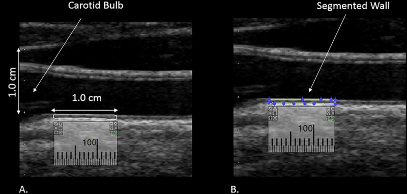
Figure 2.

Distal 1.0 cm of the common carotid artery measured to obtain the GSM value. Panel A demonstrates the calibration of 1.0 cm and the segment of the wall to be measured. Panel B demonstrates segmentation of the distal 1.0 cm of the CCA for GSM measurement.
