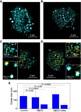
Fig. 5. Effect of lipid rafts on the nanoscale reorganization of CD44.

SR image of (A) CD44 on a methyl-β-cyclodextrin (MβCD)–treated KG1a cell that was fixed and immunolabeled by AF-647 dye in a suspension and (B) CD44 on an MβCD-treated KG1a cell that was fixed and fluorescently labeled in the microfluidic chamber after the rolling of the cell on E-selectin. Two-color SR images of (C) CD44 (cyan) and actin cytoskeleton (yellow) on an MβCD-treated KG1a cell that was fixed and immunolabeled by AF-647 dye in a suspension and (D) CD44 (cyan) and actin cytoskeleton (yellow) on an MβCD-treated KG1a cell that was fixed and fluorescently labeled in the microfluidic chamber after the rolling of the cell on E-selectin. The insets show enlarged view of the yellow regions. (E) Mean cluster sizes of CD44 on control cells, MβCD-treated cells, and MβCD-treated cells after rolling over E-selectin. Error bars indicate SDs of the cluster sizes obtained from 25, 19, and 12 cells for control, MβCD-treated, and MβCD-treated rolled cells, respectively.
