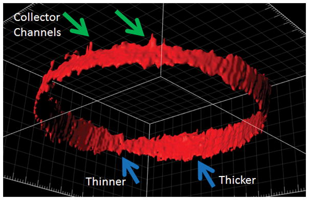
Figure 3. Circumferential (360-degree) Reconstruction of Live Human Outflow Pathways.

Anterior-segment OCT was performed circumferential around the limbus of the right eye of a healthy male. A three-dimensional AHO cast was created based on automated segmentation of Schlemm’s Canal (SC; blue arrows) and first order collector channels (green arrows). Areas of thicker and thinner SC are seen.
