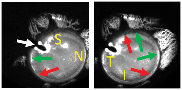
Figure 5. Aqueous Angiography During Cataract Surgery.

Aqueous angiography was performed in the right eye of a 73-year-old female during cataract surgery with 2% fluorescein diluted in balanced salt solution. The anterior chamber maintainer entered superior-temporal (white arrow) to allow space inferior-temporal for placement of an eventual phacoemulsification main wound. The subject moved her eye slightly to the right (A) and left (B) to allow viewing of different regions (S = superior, N = nasal, T = temporal, and I = inferior). Peri-limbal, there were segmental regions with (green arrows) and without (red arrow) angiographic aqueous humor outflow.
