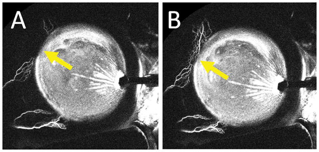
Figure 6. Dynamic Angiographic Aqueous Humor Outflow Seen by Aqueous Angiography.

Aqueous angiography was performed in the left eye of a 61-year-old female during cataract surgery with 0.4% indocyanine green diluted in balanced salt solution. The right side of the images is temporal and the left side is nasal. The anterior chamber maintainer inserted into the eye temporally. A) 176 seconds after tracer introduction and B) 182 seconds after tracer introduction. Yellow arrows show increasing angiographic signal superior-nasal over 6 seconds.
