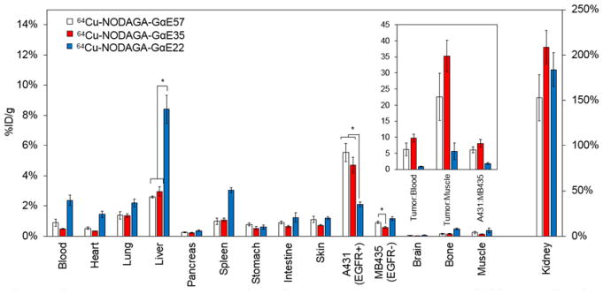
Figure 6.
Excised tissue analysis of charge modified GαE ligands. Mice xenografted with A431 (EGFRhigh) and MDA-MB-435 (EGFRlow) tumors on opposing shoulders were intravenously injected with 64Cu-NODAGA-GαE57, 64Cu-NODAGA-GαE35, or 64Cu-NODAGA-GαE22 and euthanized 2 h post-injection. Tissues or fluid of interest were extracted, weighed, and gamma decay radioactivity measured (%ID/g) (n = 5–6). *: p < 0.05. Inset represents ratios of tumor:background signal.

