Figure 1.
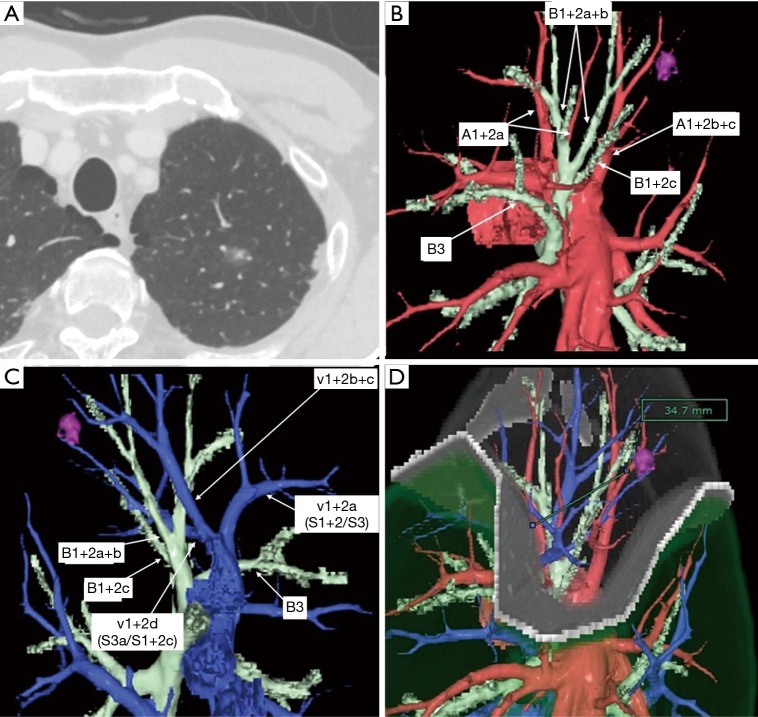
Preoperative evaluation. (A) Thin slice CT shows a part solid ground glass nodule (GGN) in the left S1+2 segment. (B) Three-dimensional (3D) CT reconstruction image shows tumor location, segmental arteries, and segmental bronchus. (C) 3D CT shows intersegmental vein. (D) Virtual intersegmental plane is made by according to the segmental artery.

