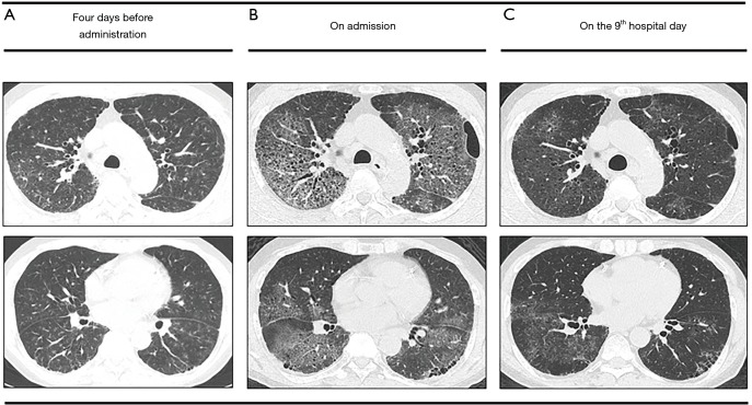
Figure 1.
High-resolution computed tomography (HRCT) of the chest. (A) Four days before administration. HRCT before administration demonstrated diffuse ground-glass opacity mainly around bronchovascular bundles with interlobular wall thickening and focal subpleural and basal reticulation with honeycombing. (B) On the day of admission. HRCT on admission demonstrated diffuse ground glass opacities with predominantly upper lobe involvement with the lesions more severe than previously noted. (C) On the 9th hospital day. Follow-up HRCT showed an improvement in the diffuse ground glass opacities overall.

